Malaria diagnostics: microscopy versus rapid tests in rural clinics
In a nationwide Kenyan school survey, malaria rapid diagnostic tests returned a sensitivity of 96.1% against expert microscopy, but a specificity of just 70.8%.
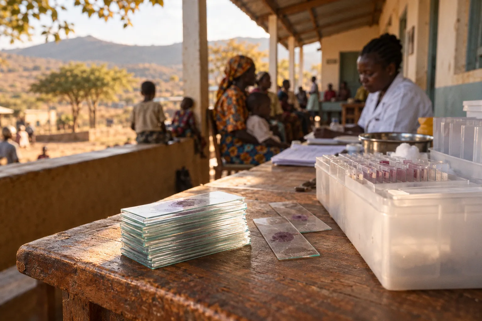
That result captures the tension facing clinical directors in Catholic mission hospitals across rural Kenya: RDTs can identify most infections under the conditions in which they were evaluated, yet they may also remain positive when a patient has recently cleared parasites or when fever has another cause. Microscopy offers information that an antigen test cannot, but it depends on stained slides, a functional microscope, reliable supplies, and trained laboratory technicians.
The choice between the two methods is therefore not theoretical. It is operational. It shapes whether a febrile patient receives antimalarial treatment, whether a persistent fever is investigated properly, and whether a rural facility can distinguish a change in malaria transmission from a change in testing practice. In a mission network where one hospital may support several smaller dispensaries, the most useful question is rarely which test should win. It is where each test can perform safely, and what happens when its result does not fit the clinical picture.
The Gold Standard: Why Microscopy Remains Essential for Species Identification
Microscopy of Giemsa-stained thick and thin blood films remains the principal reference method against which many malaria diagnostics are evaluated. It is not a flawless test, and its performance depends heavily on preparation and reader competence. When performed well, however, it provides two forms of information that are central to clinical and laboratory decision-making.
A finger-prick capillary sample is used to prepare a thick film, which concentrates blood elements and can improve the chance of detecting parasites. A thin film preserves red-cell morphology and can assist with species identification. A trained microscopist may distinguish Plasmodium falciparum from P. vivax, P. malariae, and P. ovale, although mixed infections and low parasite densities can make interpretation difficult. Microscopy can also support an estimate of parasite density per microliter of blood, a quantitative output that antigen-based RDTs do not provide.
Microscopy is valuable not because it is infallible, but because a good film can show the species and parasite burden that a binary test cannot.
For Catholic health facilities running inpatient, paediatric, and antenatal services, that additional information matters. A positive or negative line answers one question; a well-prepared film can help answer several: whether parasites are visible, which form they appear to take, whether more than one species may be present, and how substantial the parasite burden appears to be. Those observations can support the management of severe disease, the investigation of an unexpected treatment response, pregnancy-related protocols, and laboratory quality assurance.
Microscopy can also be useful during follow-up when clinicians need to assess whether parasites remain detectable. A reduction or disappearance of parasites on serial films may support a conclusion that treatment is working, but it should not be described as a direct demonstration of parasite viability or as proof of cure on its own. Film quality, timing, reader skill, prior treatment, and the possibility of low-density infection all affect the result. Clinical recovery and, where indicated, repeat assessment remain part of the interpretation.
The same caution applies to a negative thick film. It can make ongoing parasitaemia less likely when the sample was collected and examined properly, but it does not by itself definitively establish cure. A patient may have a low parasite density, an inadequately prepared film, or parasites missed during reading. In some circumstances, antigen tests may remain positive because HRP2 persists after parasites have been cleared; that persistence is a limitation of the RDT, not a reason to treat a negative film as conclusive evidence that every clinical problem has been resolved.
The laboratory is therefore more than a legacy department. It is part of the audit mechanism for the diagnostic pathway. It gives clinicians a way to investigate discordant results, monitor patterns over time, and train the next group of technicians. But its value depends on the quality of the work. A microscope sitting in a locked room, without good stain, functioning illumination, or a reader who regularly checks performance, is not the same thing as a functioning microscopy service.
Operational Realities of RDTs in Resource-Constrained Mission Settings
Rapid diagnostic tests invert the microscopy equation. A cassette, a buffer, and a small capillary-blood sample can produce a readable result in roughly 15–20 minutes. No centrifuge is required. There is no slide to stain and no microscope to align. For a rural dispensary connected to solar power but lacking a laboratory, that difference is decisive.
A community health worker or nurse can perform an RDT after focused practical training and use the result at the point of care. The test does not remove the need for clinical judgment, but it can move a facility away from treating every fever as malaria. In Catholic mission networks operating small clinics where electricity is intermittent and laboratory staffing is thin, RDTs make test-based care possible at locations that cannot sustain daily microscopy.
That operational advantage explains why RDTs have become a frontline diagnostic in many peripheral facilities across sub-Saharan Africa. A clinician at the outpatient desk does not have to send a patient home with artemisinin-based combination therapy solely because the patient is febrile. If the result is negative and there are no other indications for antimalarial treatment, the team can consider alternative causes of fever rather than treating malaria by default. If the result is positive, the treatment decision can be made promptly while the patient is still at the facility.
RDTs also simplify some aspects of logistics. Consumption can be tracked by number of tests used, while a microscopy service requires continued attention to slides, stain, buffered water, immersion oil, illumination, equipment maintenance, and staff time. That does not make RDT procurement effortless. Heat, humidity, damaged packaging, expired stock, poor storage, and inconsistent supply can all reduce the usefulness of a test that is otherwise easy to perform.
The diagnostic content is narrower than the operational convenience. Many RDTs used in malaria programmes detect histidine-rich protein II (HRP2), an antigen associated with P. falciparum, sometimes alongside a pan-malaria antigen such as pLDH. Depending on the product, the result may indicate P. falciparum or malaria more generally, but it will not provide the same species-level detail as a well-read thin film. It will not quantify parasites, and it cannot by itself distinguish a new infection from residual antigen after recent treatment.
This distinction is important in a referral network. A peripheral clinic may reasonably use an RDT to make an initial treatment decision, while a mission hospital may need microscopy to investigate persistent fever, suspected mixed infection, severe illness, or an unexpected result. The tests are answering different questions at different points in the pathway.
Navigating Diagnostic Accuracy: Sensitivity, Specificity, and HRP2 Limitations
The accuracy profile of RDTs in Kenyan field conditions is more variable than product packaging can suggest. In a study of primary school children in rural Embu District, malaria RDTs returned a sensitivity of 81.3% in 1998 and 79.3% in 2000 against blood-smear microscopy, with corresponding specificities of 81.6% and 78.3%. A broader nationwide school survey comparing three RDT brands with expert microscopy found a higher overall sensitivity of 96.1%, but a specificity of only 70.8%.
These figures are not contradictory. They illustrate how observed performance can shift with transmission intensity, parasite density, the population tested, the reference method, the product used, and the number of patients who have recently received treatment. A test result is produced by a device, but its practical accuracy is also shaped by the setting in which the device is used.
Sensitivity and specificity describe a test under particular conditions. They do not turn every positive or negative result into a complete clinical explanation.
Three limitations are especially relevant in rural Kenyan facilities.
Persistent HRP2 antigen
HRP2-based RDTs detect antigen, not living parasites. HRP2 may remain detectable after effective treatment and parasite clearance, so a positive result can persist for some time after the infection that produced it has resolved. The duration is not identical for every patient, and a positive result in someone recently treated should be interpreted alongside symptoms, treatment history, and the possibility of another cause of fever.
This is one reason a positive RDT should not automatically be treated as proof that the current fever is caused by active malaria. Microscopy may help, particularly when parasite density can be assessed, but a negative film also has to be interpreted carefully. It may fail to detect a low-density infection or be affected by poor preparation and reading. The appropriate response to discordance is investigation, not replacing one absolute rule with another.
Low-density infections and false negatives
RDTs have detection thresholds. When the parasite density is low, the amount of antigen in the sample may fall below the level required to produce a visible test line. A negative result can therefore be less reassuring when clinical suspicion is high, when the patient is at increased risk of severe disease, or when symptoms persist.
Microscopy can sometimes detect low-density infections that an RDT misses, especially when a properly concentrated thick film is examined by an experienced reader. It is not guaranteed to do so. A small sample, inadequate blood volume, poor staining, a hurried examination, or limited reader experience can also lead to a false-negative film. The practical strength of microscopy is not immunity from error; it is the additional information and the possibility of reviewing the specimen through a quality-assurance process.
pfhrp2 gene deletions
Some malaria parasites carry deletions or variations affecting the pfhrp2 gene. Where such parasites are present, an HRP2-based RDT may fail to detect a genuine P. falciparum infection because the target antigen is absent. The risk is not uniform across locations, which is why national and programme-level surveillance matters. A facility that notices repeated negative HRP2 tests in patients whose clinical picture or microscopy suggests malaria should not dismiss the pattern as random error.
The response may include checking the RDT lot, reviewing storage conditions and procedure, repeating testing through an appropriate method, and referring specimens for further investigation when available. A single negative result should be considered in context, particularly where disease is severe or the patient is deteriorating.
| Parameter | Microscopy using Giemsa films | HRP2-based RDT |
|---|---|---|
| Time to result | Often longer because films must be prepared, stained, and read | Usually about 15–20 minutes |
| Species information | Can support identification of several Plasmodium species, subject to reader skill and specimen quality | Primarily indicates the target antigen; some products also include a pan-malaria line |
| Parasite-density information | Can support an estimate of parasites per microliter | Not provided |
| Equipment | Microscope, slides, stain, buffered water, immersion oil, and dependable illumination | Test cassette, buffer, lancet or capillary device, and basic consumables |
| Operator skill | Requires trained laboratory personnel and continuing competency assessment | Can be performed by trained community health workers or nurses |
| Main limitations | Low-density infection, poor preparation, staining problems, equipment failure, and reader error | Antigen persistence, low parasite density, target-antigen deletion, storage problems, and procedural error |
| Role in a network | Detailed assessment, referral support, surveillance, and quality assurance | Frontline testing where microscopy is unavailable or too slow |
| Interpretation of a negative result | Makes detectable parasitaemia less likely but does not independently prove cure | Makes the targeted antigen less likely to be present at detectable levels; does not exclude every infection |
The table points to a practical conclusion. Neither modality should be described as structurally immune to failure. A good diagnostic system makes its failure modes visible and gives staff a route for responding to them.
Balancing Infrastructure Costs and Laboratory Staffing Requirements
Microscopy is not free. A functioning diagnostic setup requires a suitable microscope, high-quality Giemsa stain, buffered water, slides, immersion oil, cleaning materials, and a stable source of power or illumination. The equipment must be maintained. Objectives need cleaning, alignment problems need attention, and damaged microscopes need repair. A facility that receives supplies but cannot maintain its instrument may have a nominal laboratory without a dependable service.
Personnel costs are higher still. A laboratory technician trained in parasitological diagnosis represents an investment in formal education, supervised practice, competency assessment, and ongoing quality assurance. The technician must be able to prepare films consistently, recognize common species patterns, estimate parasite density, document uncertainty, and know when a specimen should be referred or re-read.
In lower-prevalence rural settings, the cost per correctly diagnosed case may rise because fixed costs are spread across a smaller number of examinations. That does not mean microscopy has lost its value. It means the service needs to be placed where it can be used often enough to maintain competence and where its additional information changes care. A mission hospital supporting several surrounding clinics may be a more sustainable microscopy hub than asking every small dispensary to maintain a separate laboratory.
RDTs have a different cost structure. The individual test is a consumable, and the infrastructure requirement is relatively small. A nurse-led outpatient team can use RDTs without a microscope room or a dedicated laboratory technician. For a peripheral Catholic mission clinic with limited referral options, that may fit the immediate budget and staffing reality better than building a full microscopy service.
But the apparent simplicity can hide other costs. RDTs must be procured continuously, stored within the manufacturer’s conditions, distributed before stock runs out, and removed when expired. Staff need supervision to ensure that the correct blood volume is applied, the right amount of buffer is used, and results are read within the specified time. Faint lines, invalid cassettes, damaged packaging, and inconsistent documentation require a response. A test that is easy to perform is not necessarily a test that is easy to govern across a dispersed network.
The cost-effectiveness question should therefore include more than the price of a cassette or the salary of a technician. A network also needs to consider:
- how many patients each facility sees and how many are likely to need malaria testing;
- whether a microscopy service can maintain staff competency at the local workload;
- the cost of referral, transport, and delayed results;
- the clinical consequences of missed low-density infections and unnecessary treatment;
- the ability to investigate persistent fever or suspected treatment failure;
- the quality of stock management and supervision;
- whether results are recorded in a form that supports surveillance and programme decisions.
Microscopy contributes to quality assurance because it offers a specimen that can be re-examined, compared, and used for training. That contribution is not automatic. Films still need clear labelling, appropriate storage, supervisory review, and a process for blinded rechecking or proficiency assessment. RDT programmes also require quality assurance, including checks on storage conditions, procedure, lot performance where appropriate, and staff interpretation.
A strategy relying exclusively on RDTs may be entirely reasonable at the first-contact level, but it narrows the network’s ability to investigate discordant results, monitor parasite density, and teach parasitology. A strategy relying exclusively on microscopy may be too slow or too resource-intensive for remote clinics. The financial decision is therefore inseparable from the clinical pathway.
Strategic Integration: Building a Hybrid Diagnostic Model for Rural Facilities
The clinical evidence does not point toward replacing microscopy with RDTs, nor toward maintaining microscopy as a monopoly. It supports a deliberate hybrid workflow. In a tiered Catholic health network, peripheral clinics can use RDTs as the frontline diagnostic because they are fast and feasible, while higher-tier facilities maintain microscopy for detailed assessment, referral support, and quantitative information.
The pathway should be explicit rather than informal. A positive RDT in an uncomplicated patient may lead to treatment according to the applicable protocol. A negative RDT in a patient with a different likely cause of fever may support a decision not to give antimalarial treatment. But when the result conflicts with the clinical picture, the protocol needs to state what happens next.
That may include referral or repeat assessment for:
- severe illness or danger signs;
- pregnancy-associated malaria;
- persistent fever after treatment;
- a positive RDT in a patient recently treated for malaria;
- a negative RDT when clinical suspicion remains high;
- suspected mixed or non-falciparum infection;
- repeated unusual results from one facility, operator, or RDT lot;
- possible treatment failure or a need to assess parasite density.
Microscopy should not be reserved only for cases that are already difficult. It also has a network-level role. A mission hospital laboratory can review selected specimens from peripheral clinics, identify patterns of discordance, support supervision, and provide a practical training platform. The value of the central service is not simply that it produces a result; it helps the network learn whether its diagnostic process is functioning.
That hybrid model requires a workforce trained in both modalities. Laboratory technicians attached to mission hospitals need continuing competency in slide preparation, staining, species recognition, parasite-density estimation, and documentation. Periodic proficiency panels, blinded rechecking, and review by a reference reader can help identify drift in performance. The exact schedule should follow the resources and applicable programme requirements, but the principle is constant: a technician who learned microscopy years ago should not be assumed to remain proficient without review.
Community health workers and nurse-led outpatient teams need a different but equally practical form of support. They should be able to:
1. collect an adequate capillary-blood sample safely;
2. perform the RDT according to the product instructions;
3. recognize a valid, invalid, positive, negative, or ambiguous result;
4. record the result and relevant recent treatment history;
5. explain when a patient needs referral or repeat evaluation;
6. avoid using a persistent positive line as automatic proof of active infection.
Lot-quality verification of new RDT consignments should be built into the network’s quality system where feasible. Storage conditions need monitoring, particularly in facilities exposed to high temperatures or irregular electricity. Stock records should be reviewed alongside testing volume and positivity patterns. A sudden fall in positive results may reflect changing transmission, but it may also indicate a supply, storage, procedural, or product problem.
The same discipline applies to microscopy. Slides need appropriate supplies, and laboratories need a system for reporting results in a way clinicians can use. A report that simply says positive or negative loses some of the method’s value. Where possible, the report should indicate the species assessment, parasite-density estimate or relevant qualifier, specimen limitations, and whether review is recommended. If the film is technically inadequate, that limitation belongs in the report rather than being hidden behind a definitive label.
The right diagnostic question is not “microscopy or RDT?” It is where each method earns its place in a defined clinical pathway. For a Catholic health network in Kenya, RDTs can extend testing to facilities that could not provide routine microscopy, while microscopy can anchor referral care, quality assurance, and investigation of difficult cases. Neither method should be treated as infallible. A positive RDT may reflect persistent antigen; a negative RDT may miss low-density infection; a negative film may reflect true absence of detectable parasites or a problem with the specimen or reading.
Rural diagnostic quality is built less by choosing one perfect test than by making the limits of every test visible to the people who use it.
The operational case is therefore clear but not simplistic. Microscopy remains essential for species-oriented assessment and parasite-density monitoring where the service can be maintained properly. RDTs remain indispensable for rapid access to testing at the periphery. Both must be staffed, supplied, supervised, and connected to a referral process that takes discordant results seriously. Diagnostic performance in rural Kenya will rise or fall on the discipline of that integration—not on the choice of cassette over microscope.